Our veterinarians are able to offer extensive medicine diagnostics work up and treatments for disease processes including the respiratory and cardiac systems, gastrointestinal system, hepatic (liver), neurologic, ophthalmologic, dermatologic, renal (kidney), haematologic (blood), endocrine/hormonal disorders and muscle disorders. Many of our veterinarians have undertaken postgraduate studies in equine medicine and surgery to assist them in their treatment of complex medical cases.
We commonly receive and treat referral medicine cases and our medicine department is an extremely important part of post surgical care in the clinic.
Respiratory Diseases
The respiratory system is one of the most common causes of poor performance in competition horses. It is second only to lameness.
Initially a history followed by a clinical examination will be undertaken. This may include the use of a rebreathing bag.
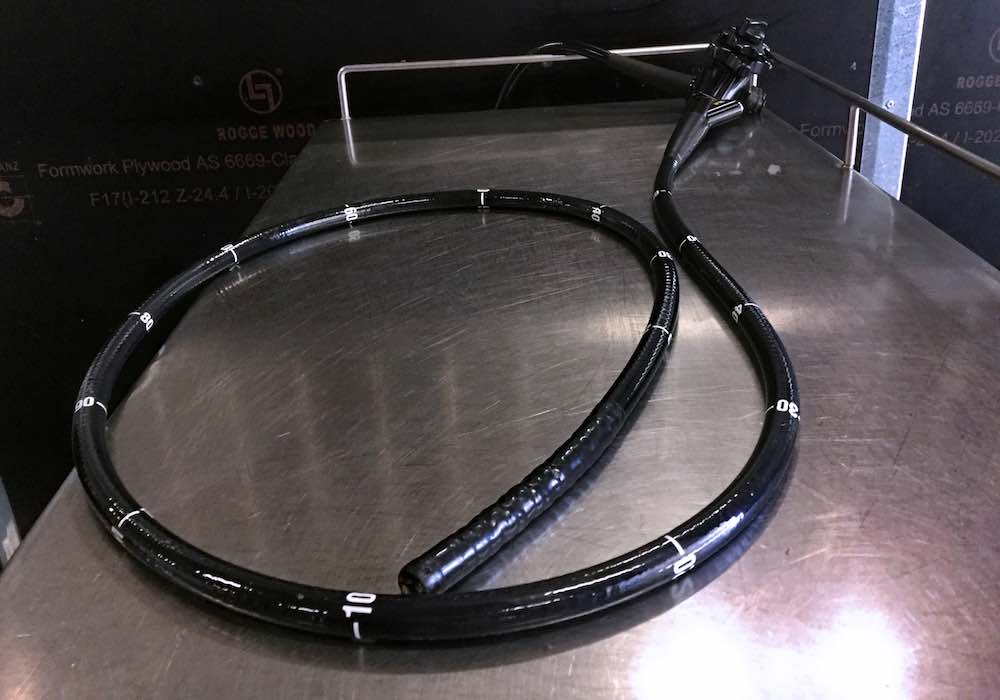
Diagnostics undertaken by our veterinarians to investigate the respiratory tract may include upper airway endoscopy, trans tracheal aspirate, bronchoalveolar lavage and thoracic ultrasound.
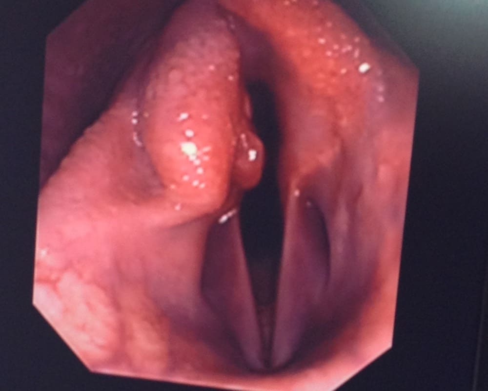
Upper airway endoscopy at rest allows the veterinarian to visualise the nasal passages, pharynx, larynx, guttural pouches, trachea and bronchi.
Resting airway endoscopy is useful in diagnosis of conditions including recurrent laryngeal neuropathy (roarers), epilglottic entrapment, ethmoid haematomas, guttural pouch infection and exercise induced pulmonary haemorrhage (EIPH bleeders).
Transendoscopic aspirate can be performed when undertaking upper airway endoscopy to sample fluid from either the trachea or guttural pouches. The tracheal and guttural fluid can be used aid in the diagnosis of infection (viral and bacterial), inflammatory, blood or neoplastic conditions.
A bronchoalvear lavage wash involves sampling the cells from the low respiratory tract by passing a long tube into the lung bronchi. It can very useful to diagnose and treat equine asthma and exercise induced pulmonary haemorrage. (EIPH)
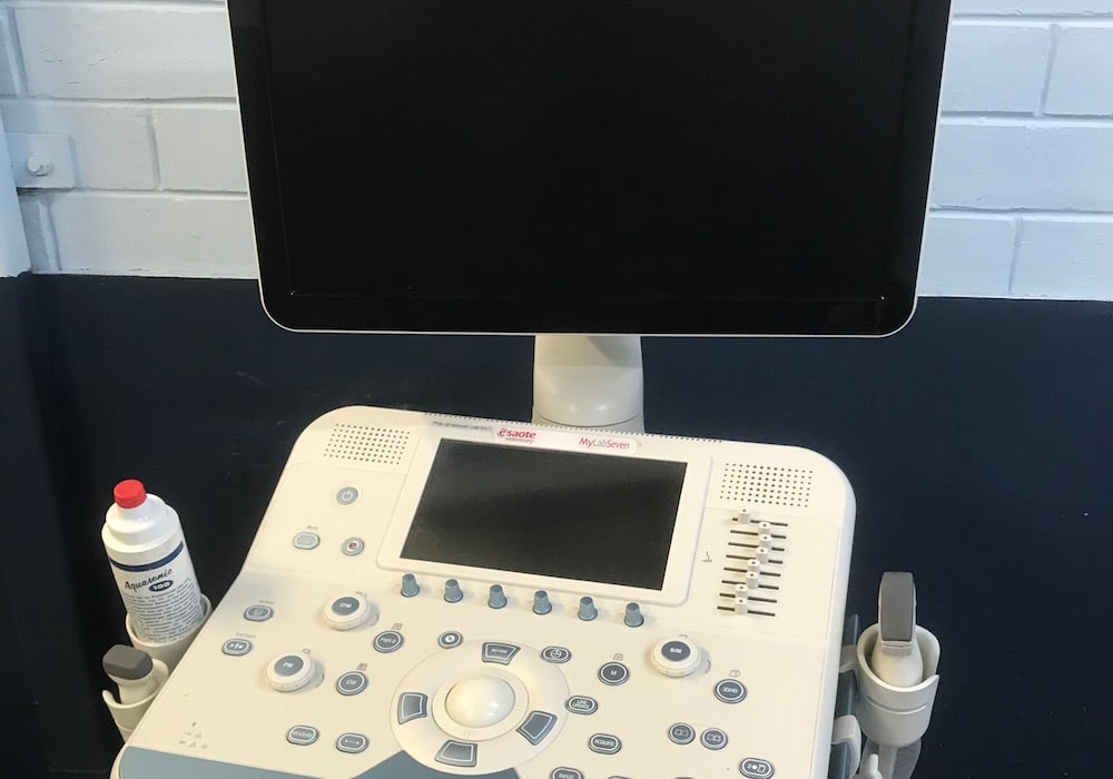
Thoracic (chest) ultrasound can be used to assess the lungs and surrounding chest cavity. Pnuemonia, neoplasia, lung and pleural abscessation can be diagnosed with chest scans. The surface of the lung can be examined and accumalation of fluid in the chest cavity can be detected. It can be useful to assist with placement of thoracic (chest) drains if required. Laryngeal ultrasound can also be undertaken to assess conditions associated with the larynx.
Overground and treadmill dynamic endoscopy are available at Morphettville equine clinic for the diagnosis of upper respiratory tract disorders in exercising horses and can be extremely useful to diagnose conditions that can not be seen at rest in both racehorses and performance horses. We have the advantage of being adjacent to the racetrack for ease of performing dynamic endoscopy.
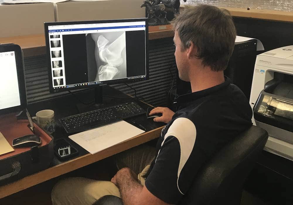
Radiography of the head is routinely performed in horses with sinus and dental disease. Radiography of the chest can also be undertaken in specific cases.

Cardiac Conditions
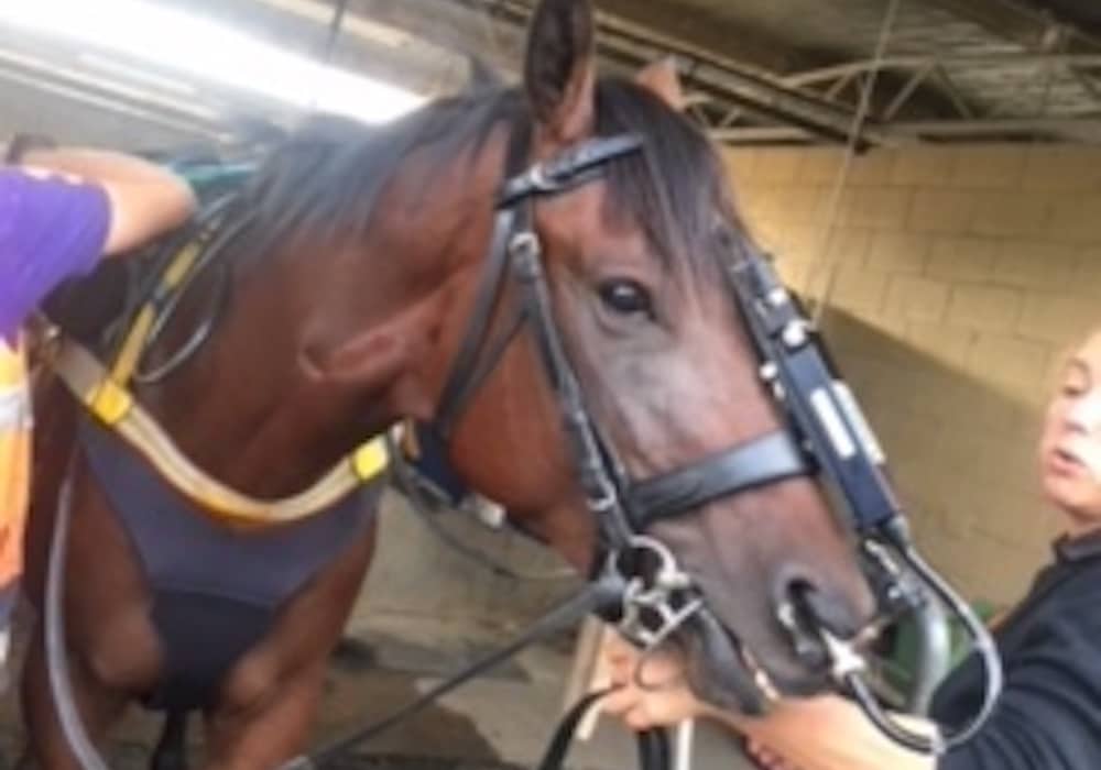
Heart abnormalities are a common finding in the horse and it is important the significance of the abnormalities can be determined.
Cardiac disease may result in heart murmurs, atrial fibrillation, exercise intolerance or poor performance.
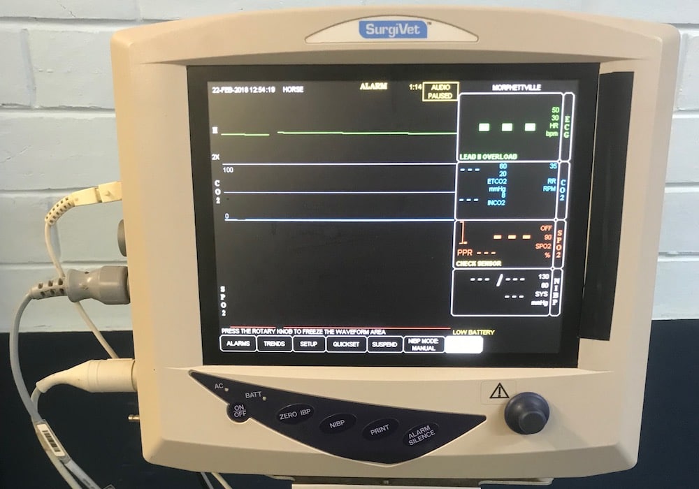
An electrocardiogram (ECG) measures the electrical activity of the heart and are commonly requested by the racing stewards following race day cardiac abnormalities.
Echocardiography (heart ultrasound) in combination with colour flow Doppler can determine the significance of heart murmurs and whether there is underlying heart disease or failure.
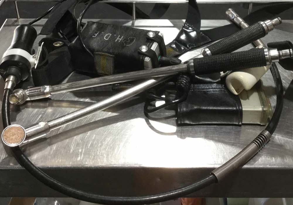
A cardiac examination at Morphettville Equine Clinic may include a general physical examination, routine blood testing, cardiac auscultation, echocardiography (heart ultrasound scan), an electrocardiogram (ECG) and cardiac troponin measurement (a measurement of cardiac muscle damage).
Heart murmurs are commonly audible when a veterinarian listens to the heart. Heart murmurs in horses are often detected during pre-purchase examination. This can lead to considerable concern over their possible future impact on the horse’s suitability for intended use. Our veterinarians may offer you a cardiac (heart) ultrasound scan to investigate a heart murmur as part of the prepurchase examination.
Gastrointestinal Disease
Gastrointestinal diseases in horses may present in a number of ways. Clinical signs can include colic, diarrhoea, inappetance, weight loss or poor performance. Certain cases can also present with a pyrexia(fever) of unknown origin but associated with the gastrointestinal system.
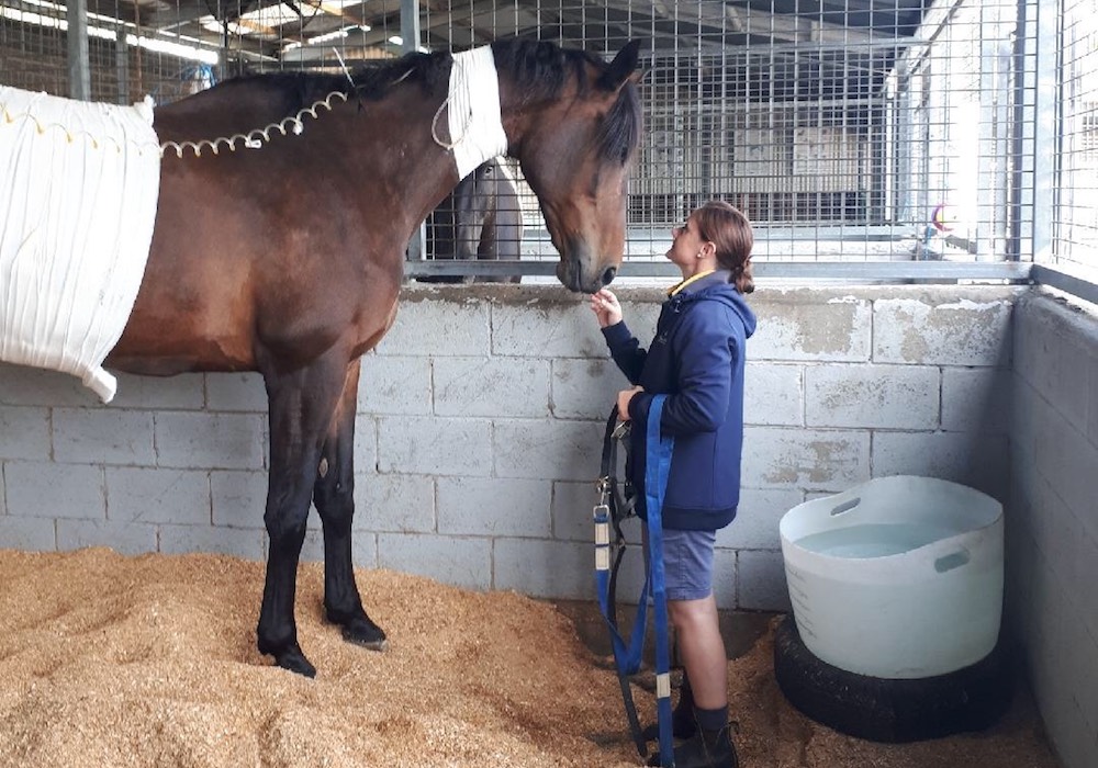
Depending on the nature of the problem, horses may be seen as day patients or admitted to the hospital in order to provide a more extensive work-up and treatment.
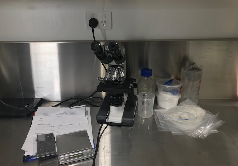
Our veterinarians regularly investigate complicated gastrointestinal disease cases. Diagnostics undertaken may include a thorough physical and dental examination, blood sampling, rectal palpation, abdominocentesis (abdominal fluid sampling), sand assessment and faecal analysis (inhouse faecal egg counts can be performed) and faecal PCR panels, gastric endoscopy, abdominal ultrasound, ventral abdominal radiography (sand assessment), dynamic glucose absorption testing and rectal mucosal biopsy.
At Morphettville equine clinic abdominal ultrasound is used routinely in the workup and assessment of colic cases. It is an important diagnostic tool in the investigation of horses with weight loss and colic signs. It can assist in the detection of peritonitis (abdominal infection), neoplasia(cancer), small intestine and large colon abnormalities and liver disease. Ultrasound guidance may be undertaken by our veterinarians to biopsy the liver or the kidneys if it clinically indicated
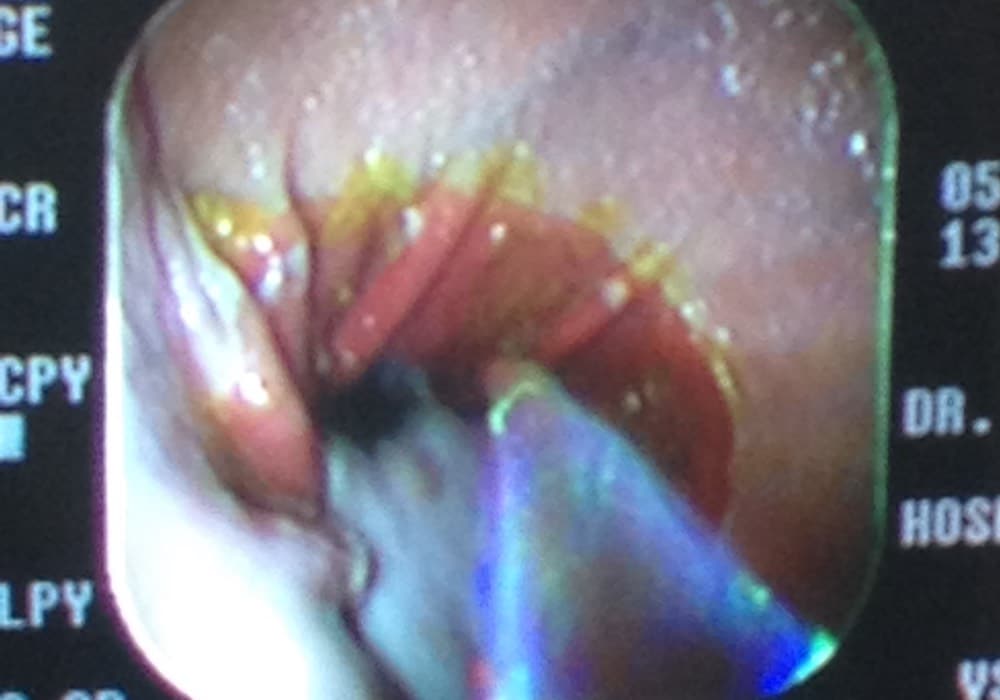
Gastrodudenoscopy allows close and detailed visualisation of many parts of the gastrointestinal tract including the oesophagus, stomach, pylorus and duodenum. Gastroscopy is the only way to diagnose gastric ulcers in horses and is extremely useful to advise of the correct treatment and management of equine gastric ulceration. Radiography of the ventral abdomen allows detection and assessment of sand.
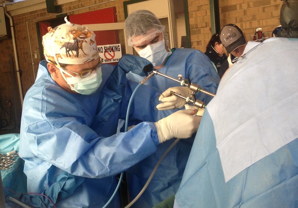
Occasionally, as part of a gastrointestinal disease investigation our surgeons will be required to perform procedures such as laparoscopy (abdomen surgery) to allow visualisation of the abdominal cavity and in some cases an extensive surgical exploration of the abdomen (exploratory laporotomy).
Neurological Disorders
Neurological diseases in the horse can be difficult to diagnose, treat, and manage. History taking is an important part of the neurological examination. Our veterinarians will undertake thorough and careful neurological examinations to determine the cause of the disease process. Diagnostics such as spinal radiography and cerebrospinal fluid analysis may also be performed.
Spinal radiography is useful to image the spine and to identify areas of possible bone damage such as osteoarthritis and spinal cord narrowing. Cervical myelography (MRI) may also be performed. This is a specialist procedure and can be conducted at HorsemedSA Morphettville by Dr Todd Booth. Cervical myelography may be required as past of the diagnosti workup of “Wobbler’s Syndrome”. Cervical facets can be radiographed and using markers, intra-articular medication administered into the spinal facets when osteoarthritis is diagnosed.
Muscular Disorders
Muscular disorders will cause a problem in horses of various disciplines from racehorses with tying up to quarter horses with muscle storeage disease. Clinical signs include exercise intolerance and poor performance. Diagnostic work-up of muscular disorder may involve blood sampling, exercise testing and muscle biopsy. Samples can be submitted to a neuromuscular laboratory in the US, which offers histopathological assessment of muscle biopsy samples to evaluate equine muscle disorders such as polysaccharide storage myopathy (PSSM). Our experience racetrack veterinarians regularly diagnosed and manage recurrent equine rhabdomyolysis (RER) in racehorses.
Ophthalmology
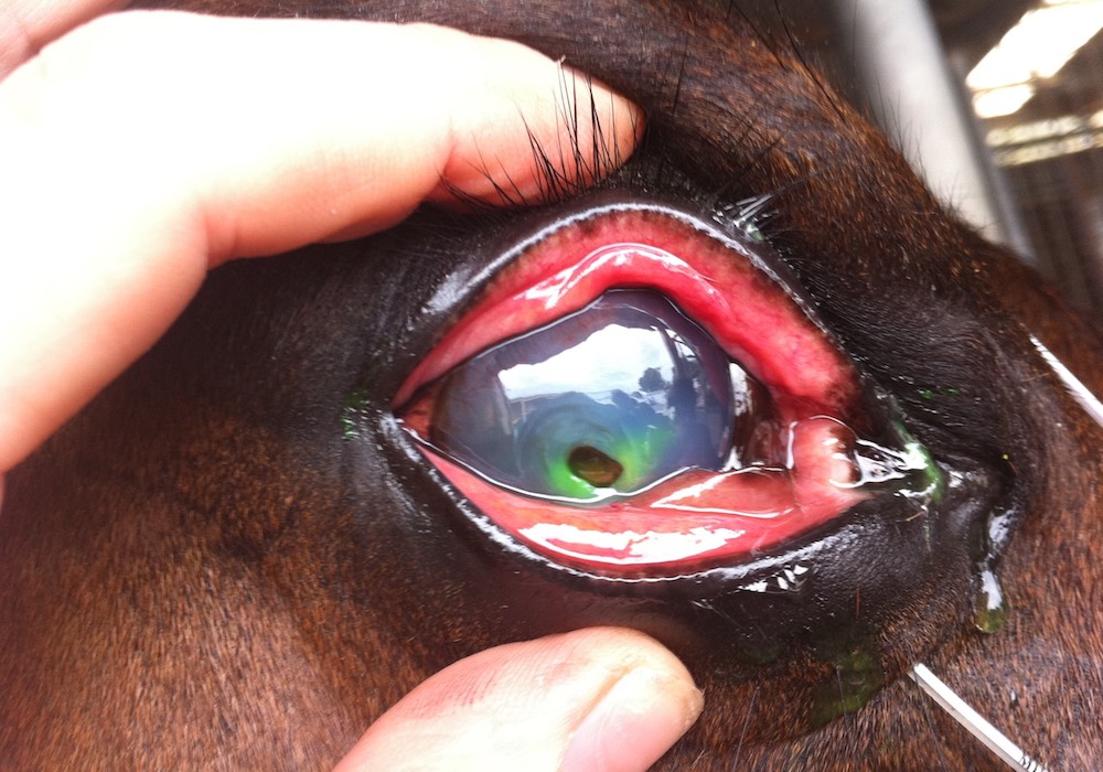
Eye injury and disease processes involving the eye are commonly seen in horses and can results in serious consequences including loss of eyesight. At Morphettville equine clinic we regularly assess complicated eye conditions with our extremely experienced clinicians. Furthermore we have the benefit of a long standing relationship with Dr. Tony Read, an ophthalmology specialist who consults at our clinic on a regular basis including performing ocular surgery.
For severe ocular disease a subpalpaebral lavage system can be placed in the eye to assist us with treating. With the advantage of intensive care, hospital based veterinarians and experienced nursing we are able to medicate the eye as frequently as required.
Endocrine Disease
Due to advancing knowledge and research in this area the diagnosis and treatment of endocrine and metabolic diseases has become a regular occurrence for our veterinarians at Morphettville Equine Clinic.
The two most common diseases we see include Equine Cushing’s Disease, and Equine metabolic syndrome (EMS). Equine Cushings Syndrome (Pars pituitary intermedia dysfunction) is a common disorder of aged horses. The disorder is caused by abnormal function of the pituitary gland. The pituitary gland is in charge of producing hormones and lies at the base of the brain. The middle lobe of the gland becomes enlarged over time, resulting in excessive hormone production.
Horses with Cushings Disease may show clinical signs such as slow shedding of the coat (hirustisM), laminitis, increased urination, increased drinking, muscle wasting, weight loss, lethargy, increased sweating, increased appetite and recurrent infections. Testing for cushing’s disease involves a simple blood test known as the ACTH blood test.
Equine metabolic syndrome (EMS) is characterised by obesity or inappropriate deposition of fat (particularly on the neck and tail base), insulin resistance (IR), and laminitis in horses and ponies. Certain breeds or individual horses are predisposed and often referred to as ‘good doers’. These horses often require less feed to maintain body weight than other horses.
EMS can be diagnosed using an oral glucose challenge test followed by measurement of insulin and glucose. It is recommended all horses are tested for both cushings diseases and EMS despite their age group.
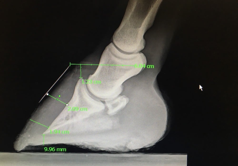
The majority of horses and ponies with cushings and EMS most commonly present with clinical signs of laminitis. At HorsemedSA, we will work closely with you and your farrier to diagnosis and treat both the laminitis and underlying disease.
Dermatology
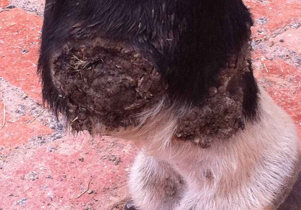
Skin diseases in horses are common and can be difficult to diagnose, treat and manage. We investigate and treat skin conditions such as itching and allergic skin reaction, nodular diseases, auto-immune skin disease and scaling and crusting. Routine diagnostic tests such as skin scrapes and biopsies may be performed.
Skin tumours such as sarcoids and squamous cell carcinomas are common in the horse. Many different treatment options, including chemotherapy and cryosurgery, are available depending on the case. We have been extremely successful treating sarcoids with the Liverpool cream (AW%) only available from the UK and imported by HorsemedSA. We have a long standing working relationship with Dr. Derek Knottenbelt in the UK regarding the treatment of extremely complicated sarcoid cases.
